


Museo di Anatomia Patologica dell'Università degli Studi di Firenze [University of Florence Museum of Pathological Anatomy]
In 1742 Antonio Cocchi, in his "Report on the Hospital of Santa Maria Nuova", noted the need to establish a Medicean Academy in Florence. Finally in 1824, after various attempts, the Florentine Medical-Physical Society was founded, thanks also to Giovan Pietro Vieusseux and his Cabinet. The members of the Society proposed to study the medical sciences through strictly experimental methods. Among the most active and famous members during these early years were Pietro Betti, Maurizio Bufalini and Filippo Pacini. The latter, in 1835, made a report to the members in which he described the discovery of the tactile corpuscles, denominated in 1844 the "Pacini corpuscles". Particularly interesting was the vivacious debate on the nature of cholera, which served to promote studies on rules of hygiene and drinking water. In 1886 the Society assumed its present name of Florentine Medical-Physical Academy.
In the years of its foundation the society decided, especially at the urging its president Pietro Betti, to set up a Pathological Museum. The Museum took on a central role in the Tuscan medical panorama thanks to a Royal Decree of January 28, 1839, which prescribed its mansions. Regulations for conducting autopsies in the Hospital of Santa Maria Nuova were established by the decree. Each autopsy was to be presided over by the director of the Pathological Museum. The deceased patient's clinical history was to be read and filed. The diagnosis made by the patient's doctor was to be compared with the results of the autopsy. The pathological organs, removed by surgical procedures, were to be consigned to the Museum. In cases where patients were cured, their doctors were required to send the Museum a report on their post-operatory care.
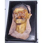
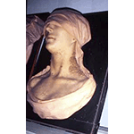
Due to the difficulty of ensuring correct conservation of the pathological materials, it was decided to have some duplicates fabricated in wax. The Museum's model-makers referred to the experience and the techniques practised in the other wax-modelling laboratory in Florence, that of the Specola Museum. Many models, often surprisingly realistic, were fabricated, providing a fascinating glimpse of the major pathologies in the 19th century. The collection of anatomical wax figures includes numerous reproductions, mainly the work of Giuseppe Ricci, Luigi Calamai and Egisto Tortori. Noteworthy among those attributed to Calamai is the so-called "leper" (1851). The modellers of the earliest works are unknown. A remarkable example of symbiosis between science and art, the wax models were important above all for their value in teaching, allowing professors to illustrate the most important diseases to future physicians without having to recur to the dissection of cadavers. The Museum, which attracted illustrious researchers in European medicine, has a small anatomical collection that includes organic remains, such as skeletons and parts of organs. In 1840, also consequent to the Museum's activity, a chair of pathological anatomy was instituted at the Hospital of Santa Maria Nuova.
The Institute of Pathological Anatomy and the Museum were moved to Careggi in 1959. At present the Museum's collections are managed by the Department of Human Pathology and Oncology, instituted in 2000.
****************************
Texts by Graziano Magrini
English translation by Catherine Frost
Last update 11/feb/2008