

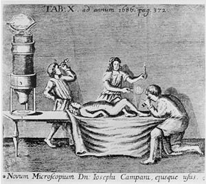
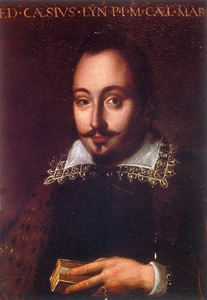
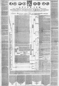
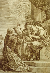
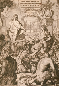
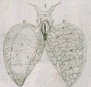
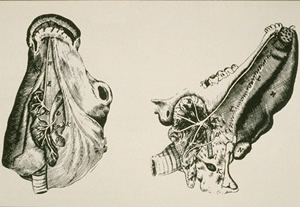
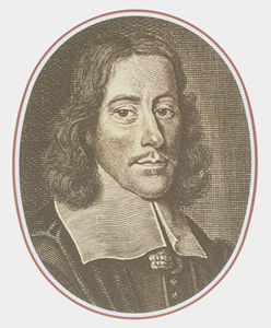
Microscopic anatomy (fig.1) began during the course of the 17th century with Federico Cesi (1585-1630) (fig.2) and Francesco Stelluti (1577-1651) in the Apiarium (Rome, 1625) (fig.3), a work covering a single folio of extraordinary size, containing detailed descriptions of naturalist, historical-erudite and literary nature on bees.
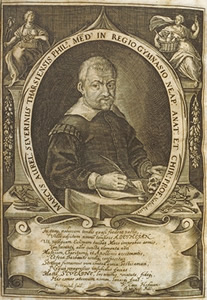
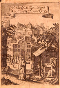
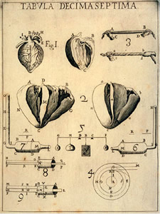
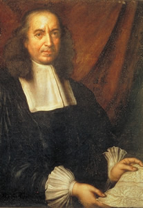
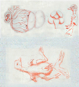
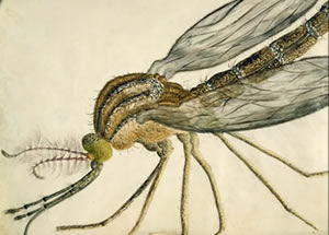
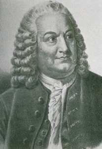
Later, Giovanni Battista Hodierna (1597-1660) published, in L'occhio della mosca (Palermo, 1644) (fig.4), a text dedicated to the anatomy of insects, a masterly example of naturalist research conducted with the aid of the microscope; Marco Aurelio Severino (1580-1656) (fig.5) in his Zootomia Democritaea (Nuremberg, 1645), justly considered the first treatise on comparative animal anatomy, proposed an atomistic conception of animal structures developed on the basis of microscopic observation. (fig.6)
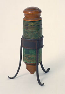
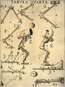
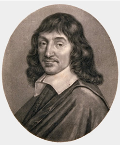
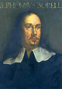
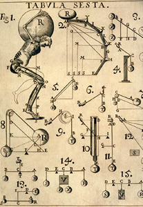
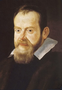
Gradually, the microscope (fig.7) helped to disclose the causes of the functioning of organisms (fig.8) which were explained by extending to the biological sphere the rigorous style (fig.9) of geometric analysis employed by Galileo (1564-1642) in studies on mechanics. This aspect was developed especially by René Descartes (1596-1650) (fig.10) and by Giovanni Alfonso Borelli (1608-1679) (fig.11). The latter, in particular, described in a mechanical perspective the muscular movements involved in walking, running (fig.12), and lifting weights (fig.13) as well as the internal motions of the body.
Microscopic anatomy was however developed in all of its potentiality by Marcello Malpighi (1628-1694) (fig.14). As Galileo (fig.15) had launched exploration of the great machine of the universe with the telescope (fig.16), so Malpighi aimed to reveal the hidden structure of the machine that was the human body with the microscope (fig.17). He observed the alveolar structure of the lungs (fig.18), the papillary receptors on the tongue (fig.19), the connection between arterial and venous blood vessels (fig.20), identified the red blood cells and described precisely the first stages in the embryonic development of a baby chick. (fig.21).
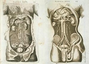
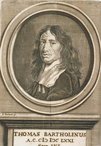
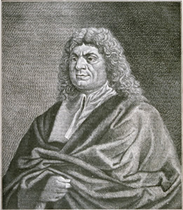
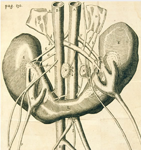
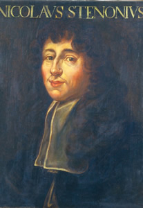
The combination of "thin" anatomy and microscopic magnification soon led to a succession of remarkable discoveries (fig.22) Thomas Bartholin (1616-1680) (fig.23) identified the lymphatic ducts (fig.24); Lorenzo Bellini (1643-1704) (fig.25) revealed the structure and function of the kidneys (fig.26), furnishing an explanation of the mechanical type; Francesco Redi (1626-1697) (fig.27) illustrated the extraordinarily complex organization of insect life (fig.28); Thomas Wharton (1614-1673) formulated the theory of the glands as secretory organs (fig.29); Niels Steensen (1638-1686) (fig.30) conducted accurate microscopic observations of muscle fibers; Thomas Willis (1621-1675) (fig.31) e poi Albrecht von Haller (1708-1777) (fig.32) studied the structure of the nervous system and the dynamics of neuro-muscular functions (fig.33).


Click on the image to enlarge it
| ©2007 IMSS · Piazza dei Giudici 1 · 50122 Florence · ITALY |